Ear Diagrams 21
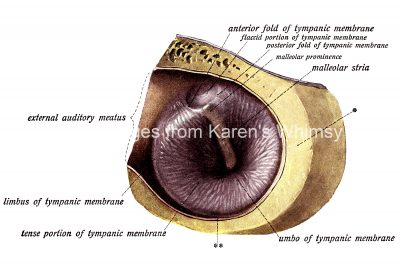
The right tympanic membrane as seen from the outer side. (Enlarged five times.)
The grater portion of the wall of the external auditory meatus has been removed. * = Cut surface of the bone. ** = Cross-section of the mucous membrane of the external auditory meatus.