Ear Diagrams 17
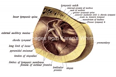
The outer wall of the right tympanic cavity as seen from the outer side. (Enlarged five times.) The tympanic membrane has been removed together with the wall of the external auditory meatus. * = Layer of periosteum which conceals the tensor tympani.